Cells, like all of us, live in a matrix. And the nature of the
Cells live in a microenvironment with a mechanical component which regulates their behavior. We have explored how the mechanical stimuli in the extracellular space regulate various cell phenotypes, with a particular focus on the cardiovascular milieu. We have developed methods to mature human iPSC derived cardiomyocytes to an adult-like state, and develop in vitro substrates for drug screening.
In recent years, inspired by the evolutionary changes we have observed across mammals, we have begun to explore how the altered mechanics in the scar tissue inform epithelial invasion. We have used cesarean scar as a “common and standard model” of scar to study how the changed mechanics can result in invasive pathology, e.g. placenta accreta.
Key Findings
- Although human iPSCs (or ESCs) can be efficiently differentiated into cardiomyocytes, these protocols only produce “fetal like” cells. iPSC derived cardiomyocytes (iPSC-CMs) are a great hope because they can serve as surrogates for essential drug screenings, as it is widely recognized that small animal models for cardiotoxicity are not accurate. How do we address this challenge? We have developed CMM: Cardiac Mimetic Matrix to accelerate maturation of differentiated human iPSC-CMs into a more adult-like state in a “physiological trajectory”. Our state of the art system is particular suited for metabolic maturation of these cells, setting critical standards for differentiated cardiomyocytes (Afzal J. et al., Cell Reports, 2022).

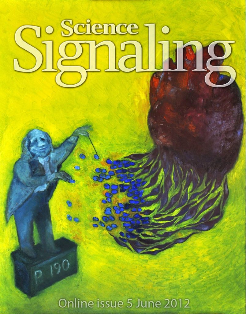
- The mechanical microenvironment can orchestrate multiple signaling pathways to choreograph a complex process of tissue development and morphogenesis. We had shown that p190RhoGAP, a GAP protein for small Rho GTPase, can act as an adapter protein to convey the extracellular mechanical signals in the heart to (i) selectively proliferate endothelial progenitors, (ii) differentiate progenitors towards endothelial state, (iii) morphogenetically organize them into luminal structures —– all in different time scales by regulating multiple different temporally dynamic pathways (Kshitiz et al., Science Sig, 2012).

Relevant Publications
- Du W, Novin A, Liu Y, Afzal J, Suhail Y, Gavin NR, Jorgensen J, Schmidt T, Morosky CM, Sanders M, Brewer M, Kshitiz (2024), Scar matrix drives Piezo1 mediated stromal inflammation leading to placenta accreta spectrum, Nature Communications, 8379.
- Du W, Novin A, Liu Y, Afzal J, Liu S, Suhail Y, Kshitiz (2024), Oscillatory hypoxia induced gene expression predicts low survival in human breast cancer patients, Mechanobiology in Medicine 3, 100070-11.
- Afzal J, Kshitiz (2023), CRISPRing the hypertrophic cardiomyopathy: correcting one pathogenic variant at a time, Signal Transduction & Targeted Therapy, Nature Publications 116(254).
- Afzal J*, Du W*, Novin A, Liu Y, Wagner G, Kshitiz (2022), Paracrine HB-EGF signaling reduce enhanced contractile and energetic state of activated decidual fibroblasts by rebalancing SRF-MRTF-TCF transcriptional axis, Frontiers in Cell & Development Biology, 10, 927631.
- Afzal J±, Liu Y, Du W*, Suhail Y*, Zong P, Feng J, Ajeti V, Sayyad WA, Nikolaus J, Yankova M, Deymier AC, Yue L, Kshitiz* (2022), Cardiac ultrastructure inspired matrix induces advanced metabolic and functional maturation of differentiated human cardiomyocytes, Cell Reports, 40(4), 111146.
- Suhail Y, Cain MP, Vanaja K, Kurywchak PA, Levchekno A, Kalluri R, Kshitiz (2019). Systems Biology of Cancer Metastasis, Cell Systems, 9(2), 109-127.
- Park JS, Kim DH, Shah SR, Kim HN, Kshitiz, Kim P, Quinones-Hinojosa A, Levchenko A (2019). Switch-like enhancement of epithelial-mesenchymal transition by YAP through feedback regulation nof WT1 and Rho-family GTPases, Nature Communications, 2797.
- Kim DH, Ewald AJ, Park JS, Kshitiz, Kwark M, Gray RS, Su CY, Seo J, An SS, Levchenko A (2018), Biomechanical interplay between anisotropic re-organization of cell and the surrounding matrix underlies transition to invasive cancer spread, Scientific Reports, 14210.
- Suhail Y, Cain MP, Vanaja K, Kurywchak PA, Levchekno A, Kalluri R, Kshitiz (2019). Systems Biology of Cancer Metastasis, Cell Systems, 9(2), 109-127.
- Kshitiz, Afzal J, Chang H, Goyal R, Levchenko A (2015). Mechanics of Microenvironment as Instructive Cues Guiding Stem Cell Behavior, Current Stem Cell Biology, 2(1), 62-72.
- Kshitiz, Afzal J, Kim SY, Kim DH (2015). A nanotopography approach for studying the structure-function relationships of cells and tissues, Cell Adhesion & Migration, 9(4), 300-307.
- Kshitiz, Afzal J, Kim DH, Levchenko A (2014). Mechanotransduction via p190RhoGAP regulates a switch between cardiomyogenic and endothelial lineages in adult cardiac progenitors, Stem Cells, 32(8): 1999 2007.
- Ahn EH*, Kim Y*, Kshitiz*, An SS, Afzal J, Lee S, Kwak M, Suh KY, Kim DH, Levchenko A (2014). Spatial control of adult stem cell fate using nanotopographic cues, Biomaterials, 35(8):2401-10.
- Suhail Y*, Kshitiz*, Lee J, Walker M, Kim DH, Brennan MD, Bader JS, Levchenko A (2013). Modeling intercellular transfer of biomolecules through tunneling nanotubes, Bulletin of Mathematical Biology, 75(8):1400-16.
- Kim DH*, Kshitiz*, Smith R, Kim P, Marban E, Suh KY, Levchenko A, Nanopatterned Cardiac Cell Patches Promote Stem Cell Niche Formation and Myocardial Regeneration, Integrative Biology, 4(9):1019-33. (cover article)
- Kshitiz, Park JS, Kim P, Helen W, Engler AJ, Levchenko A, Kim DH. (2012). Control of stem cell fate and function by engineering physical microenvironment, Integrative Biology, 4:1008-1018.
- Kshitiz, Kim DH, Beebe D, Levchenko A. (2011). Micro- and nanoengineering for stem cell biology: the promise with a caution, Trends in Biotech, 29(8):399-408.
- Niu X, Gupta K, Yang JT, Shamblott MJ, Levchenko A. (2009). Physical transfer of membrane and cytoplasmic components as a general mechanism of cell-cell communication, J Cell Sci., 122(Pt 5):600-610.
